Fig. 2.
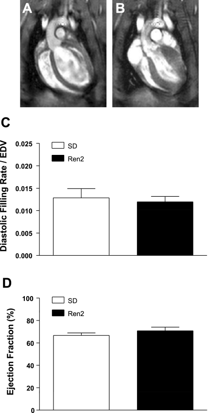
In vivo cine MRI analysis of left ventricular (LV) function in SD and Ren2 rats. Nine-week-old male Ren2 and SD rats were imaged by in vivo cine MRI. A and B: representative images of end systole (A) and end diastole (B) of a representative SD rat are shown. Movies for a representative SD and Ren2 rat are shown in Supplemental Fig. 1. C: no differences or similarities were found in the diastolic filling rate/end-diastolic volume (EDV). D: the ejection fraction between SD and Ren2 rats was significantly similar (power = 0.842) via a noninferiority test.

