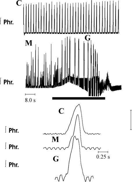
Fig. 6.
Activity of the phrenic nerve in eupnea, before and after administration of methysergide, and in gasping in a wild-type mouse. C represents traces obtained in eupnea. Note incrementing pattern of incrementing phrenic discharge. M represents recordings 10 min after addition of 2 μM methysergide to the perfusate. Frequency of phrenic bursts had increased and peak height was variable. Solid horizontal bar shows period of ischemia, during which perfusion was terminated. Pattern of phrenic discharge was altered to the decrementing pattern of gasping (G). On a recommencement of perfusion, rhythmic phrenic discharge did not return for 30 s.

