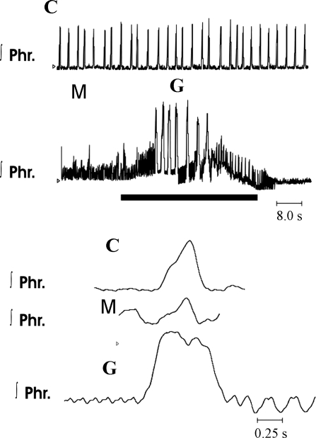
Fig. 8.
Alterations in integrated phrenic discharge after administration of methysergide to a Pet-1 homozygous mouse. C represents control recordings; M represents recordings obtained 5 min after addition of 3 μM methysergide to perfusate. Note increase in frequency and decrease in peak height of integrated phrenic bursts. G represents gasps induced during period of ischemia (solid horizontal bar). Rhythmic phrenic bursts did not return until 116 s after recommencement of perfusions. Bottom traces are on a faster time scale than top traces.

