
Figure 1. Similar percentage and frequency of different splenic cDCs populations, macrophages and granulocytes in spleens of WT, IFN-β-/- and IFNAR-/- mice.
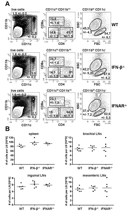
(A) Splenocytes from WT, IFN-β-/- and IFNAR-/- were isolated, stained for following markers: CD11c, CD11b, CD4, CD8α and B220 and analyzed by flow cytometry. Data are representative of three independent experiments with at least 5 mice per group. (B) Similar number of leukocytes per lymph node or spleen in WT, IFN-β-/- and IFNAR-/- mice.