
Figure 6. Splenic cDCs from IFN-β-/- and IFNAR-/- mice form lower levels of MHC I/SIINFEKL complexes.
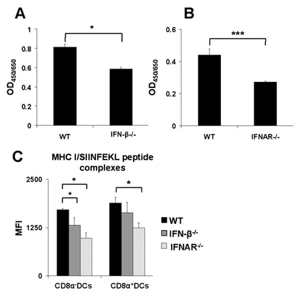
Splenic cDCs were sorted out from spleens of WT, IFN-β-/- or IFNAR-/- mice, pulsed with SIINFEKL peptide (OVA257-264) for 1h, washed intensively and (A, B) co-cultured with the SIINFEKL/H-2Kb restricted B3Z hybridoma T cells for 24h. Cells were then lysed and monitored for LacZ expression by the introduction of ONPG substrate. Optical density was measured at 450 nm with wavelength correction set at 650 nm. (C) cDCs were stained with 25-D1.16 antibody, MFI was measured by flow cytometry, graphs show values for 100 ng/ml of SIINFEKL peptide, cells were gated on CD8α+DCs and myeloid DCs (CD8α-). Results are representative of at least three mice for WT, IFN-β-/- and IFNAR-/- in three independent experiments. Statistical significance was determined using the paired Student's t test. * P<0.05; *** P<0.005