
Figure 8. In vivo inhibition of Hsp70 by 15-deoxyspergualin or knockout of Hsp70.1 and Hsp70.3 leads to impaired antigen presentation by splenic cDCs.
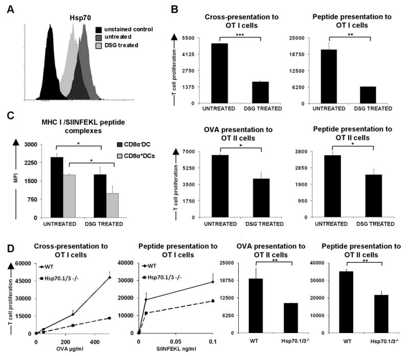
(A) Intracellular staining of Hsp70 in splenic cDC from mice untreated and treated with 15-deoxyspergualin. (B) Antigen presentation assay with splenic cDCs from untreated mice and mice treated with 15-deoxyspergualin. Splenic cDCs were incubated with purified, CFDA(SE) labeled OT I or OTII cells for 1.5 days (class I restricted peptide) and 2.5 days. OVA257-264 peptide concentration 10 ng/ml, OVA323-339 2 μg/ml, OVA protein 1mg/ml (class II presentation) or 500 μg/ml (class I presentation). The proliferative response of T cells was enumerated by flow cytometry. Data are representative of at least five mice for WT untreated and DSG treated in three independent experiments. (C) DCs from untreated and DSG treated mice were loaded for 1h with 100 ng/ml of SIINFEKL peptide, washed and stained with 25-D1.16 antibody recognizing MHC I/SIINFEKL peptide complexes. Graph shows MFI values for five mice per group. Data are representative of two independent experiments. (D) Antigen presentation assay with splenic cDCs sorted from Hsp70.1/3-/- and WT mice. Cells were loaded with appropriate OVA derived peptides or whole OVA, washed and incubated in vitro with CFDA(SE) labeled OT I or OTII cells for 1.5 days (class I restricted peptide) and 2.5 days. OVA323-339 peptide concentration 2 μg/ml, OVA protein 1mg/ml (MHC II presentation). The proliferative response of T cells was enumerated by flow cytometry. Data are representative of 3 mice for Hsp70.1/3-/- and WT in two independent experiments. Statistical significance was determined using the paired Student's t test. * P<0.05; ** P<0.01; *** P<0.005