
Figure 4.
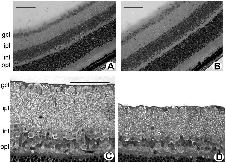
Histology of retinas of cbs+/− mice (age, 3 weeks and 1–2 years). Light micrographs of hematoxylin-eosin-stained cryosections of retinas of 3-week-old wild-type (cbs+/+) mice (A) and heterozygous (cbs+/−) mice (B). Light micrograph of toluidine blue–stained, plastic-embedded retinas of 1-year-old wild-type (cbs+/+) mice (C) and heterozygous (cbs+/−) mice (D). Scale bar, 50 μm.