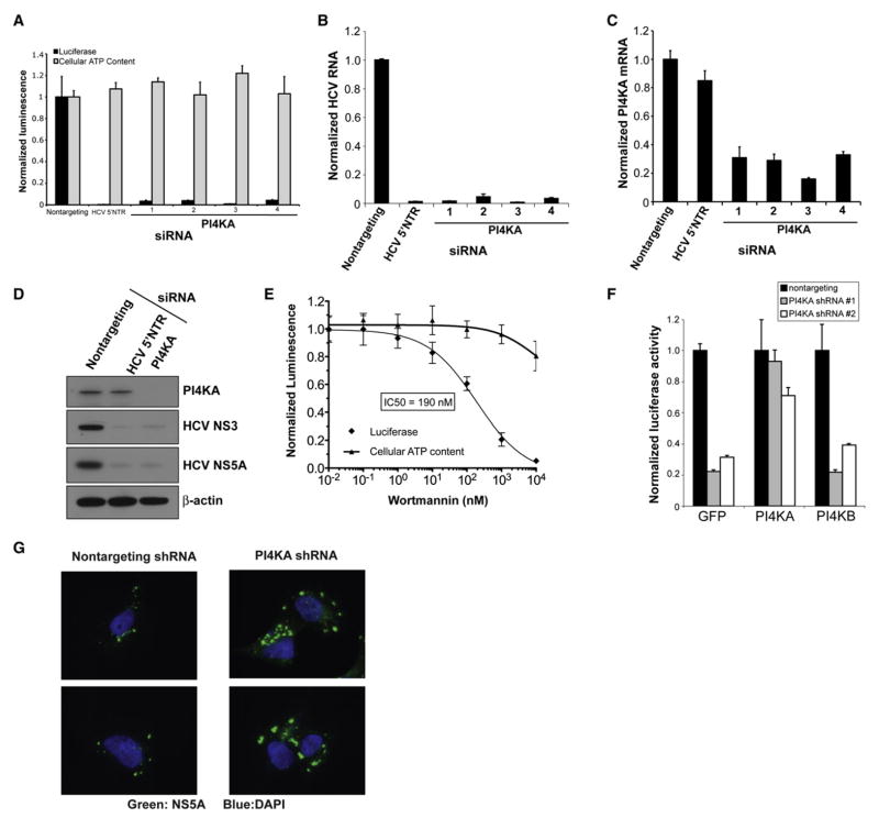
Figure 2. PI4KA Is Essential for Hepatitis C Virus Replication.
(A) PI4KA silencing with four individual siRNA duplexes blocks replication of the full-length HCV replicon OR6 (black bars) without significant cytotoxicity as measured by cellular ATP content (gray bars). Values were obtained from quadruplicate wells in two independent experiments and are mean ± SD.
(B) siRNAs against PI4KA block replication of the infectious JFH1 HCV strain. Cells were infected with JFH1 48 hr posttransfection, and HCV RNA was quantified by quantitative PCR (qPCR) at 24 hr postinfection. Values were obtained from triplicate qPCR replicates from duplicate wells and are mean ± SD.
(C) The magnitude of HCV inhibition parallels the degree of PI4KA silencing. PI4KA transcripts were quantified by qPCR 48 hr posttransfection. Values are mean± SD.
(D) PI4KA silencing depletes HCV nonstructural proteins in OR6 replicon cells as determined by immunoblotting with the indicated antibodies. Cell lysates were prepared from OR6 cells transfected with the indicated siRNAs for 72 hr.
(E) Wortmannin inhibits HCV replication with an IC50 consistent with a type III PI 4-kinase. OR6 replicon cells were treated with the indicated concentrations of wortmannin for 24 hr. Replicate wells were assayed for luciferase activity (diamonds) or cellular ATP content (triangles). Values were obtained from quadruplicate wells in two independent experiments and are mean ± SD.
(F) PI4KA shRNAs block HCV replication and can be rescued by an RNAi-resistant PI4KA construct but not by PI4KB. OR6 replicon cells were transduced with MMLV retroviral vectors encoding GFP, PI4KA, or PI4KB cDNAs lacking the 3′UTR. The cells were then transduced 24 hr later with lentiviral vectors encoding a nontargeting shRNA (black bars) or two different shRNAs targeting the PI4KA 3′UTR (gray and white bars). Cells were assayed 96 hr after lentiviral transduction. Values were obtained from quadruplicate wells in two independent experiments and are mean ± SD.
(G) PI4KA silencing induces the formation of abnormally large NS5A-positive membrane structures. UHCVcon57.3 cells were transduced with the indicated shRNA constructs 4 days prior to induction of HCV polyprotein expression by withdrawal of tetracycline from the growth medium. Twenty-four hours after induction, cells were fixed and stained with a monoclonal antibody to HCV NS5A (green) and counterstained with DAPI to highlight cell nuclei (blue).

