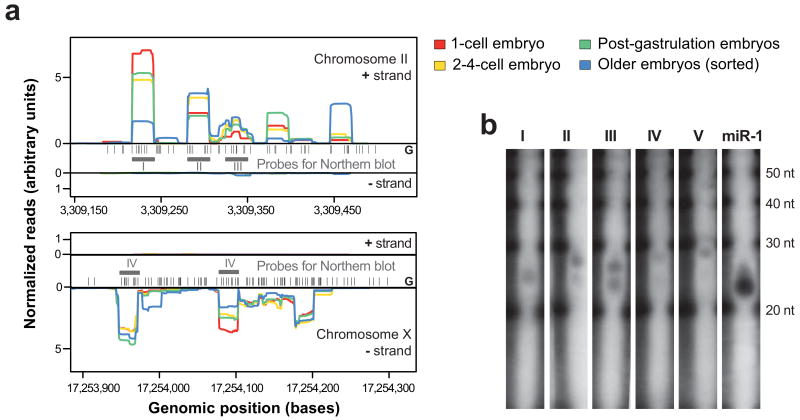
Figure 6.
26G-RNAs are expressed from intergenic clusters
(a) Two genomic clusters of 26G-RNAs on chromosome II and chromosome X. We observed most of the reads from one strand in our embryo samples. Black bars represent the positions of 5′ Guanine nucleotides in the genome. Grey bars and roman numerals indicate the features for which Northern blots were performed. Profiles are normalized to yield identical areas under the sense strand curves. (b) Validation of five (out of five tested) 26G-RNAs by Northern blots with total RNA from mixed embryos. The observed transcript lengths vary slightly between 24-28 nt. Some of the tested probes exhibit a double band in the Northern blots (II, III, IV), while others (I and V) exhibit only a single band. A 21 nt probe for miR-1 was used as a positive control.

