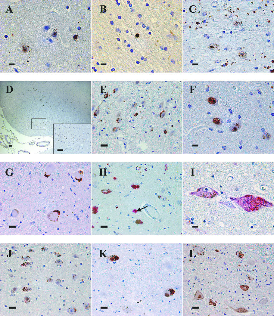
Figure 4.

Patterns of 1C2 immunoreactivity in the CNS of HD cases. (A) Frontal cortex of Case 2 demonstrating types of immunoreactivity: coarse granules in neuropil, finely granular aggregated staining in neuronal cytoplasm, diffuse nuclear staining, and round and oval aggregated nuclear immunoreactivity. (B) Subcortical white matter with a diffusely immunoreactive glial nucleus in Case 11. (C) Caudate nucleus of Case 1 with intranuclear immunoreactivity and abundant granular neuropil staining. (D) Entorhinal cortex of Case 5 showing immunoreactive layer 2 pre-alpha clusters. (E) Lateral geniculate body of Case 3 displaying multiple neurons with aggregated cytoplasmic immunoreactivity. (F) Thalamus of Case 2 demonstrating nuclear and cytoplasmic immunoreactivity. (G) Aggregated cytoplasmic immunoreactivity of dentate nucleus neurons of Case 11. (H) Substantia nigra neurons containing aggregated cytoplasmic immunoreactivity (red) intermixed with brown neuromelanin in Case 2. Some neurons have neuromelanin alone. Aggregated nuclear immunoreactivity (arrow) is present in one neuron. (I) Two neurons of the locus coeruleus of Case 15 demonstrating diffuse and aggregated cytoplasmic immunoreactivity, one also with faint, diffuse nuclear immunoreactivity. (J) Basis pontis of Case 2 with frequent immunoreactive neurons. (K) Reticular formation neurons in medulla of Case 5 with intense aggregated cytoplasmic staining and diffuse and aggregated nuclear immunoreactivity. (L) Nucleus dorsalis of thoracic spinal cord of Case 5 showing diffuse and aggregated cytoplasmic immunoreactivity. Several neurons have diffusely immunoreactive nuclei. Scale bars: A, B, C, F, I = 10 µm; E, G, H, J, K, L = 25 µm; D and inset = 100 µm.
