FIG. 3.

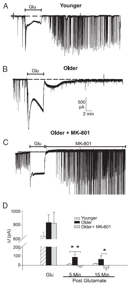
Glutamate exposure induces persistent inward current that is dependent on age and NMDA receptor. A–C: representative examples of spontaneous excitatory postsynaptic current (EPSC) activity and inward current in neurons voltage-clamped at −80 mV, recorded before, during, and after a 5-min application of 20 μM glutamate (Glu). Dashed line shows baseline holding current (Ih). In Younger neurons (n = 25) (A), glutamate application resulted in a large increase in inward current followed by rapid restoration of baseline Ih after glutamate termination. B: Older neurons (n = 14) exhibited a somewhat larger (but variable and nonsignificant) increase in inward current during glutamate exposure. After glutamate exposure, holding current remained elevated above preinsult levels for >5 min before eventually recovering. Postinsult period in Older neurons was also characterized by an increase in the appearance of smaller EPSCs and an almost complete loss of larger (>300 pA) EPSCs. C: postinsult increase in inward holding current and changes in EPSC activity were completely reversed in Older neurons by applying MK-801 (10 μM) immediately after the glutamate insult (n = 9). D: mean ± SE change in holding current (ΔI) during glutamate exposure and at 5 and 15 min after glutamate application (relative to preinsult levels) in Younger (white columns), Older (filled columns), and Older MK-801–treated (gray columns) neurons. *P < 0.05.
