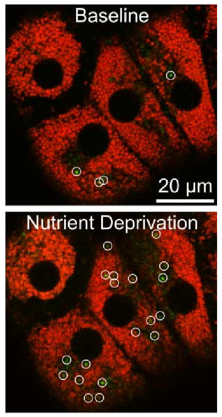
Figure 3. Mitochondrial Depolarization in Hepatocytes after Nutrient Deprivation.

Cultured rat hepatocytes were loaded with MitoTracker Green and TMRM and imaged by confocal microscopy before (Baseline) and 60 min after (Nutrient Deprivation) changing from complete growth medium to a modified Krebs-Ringer buffer containing glucagon. Green-fluorescing structures (circles) in the overlay images are newly depolarized mitochondria.
