Figure 4.
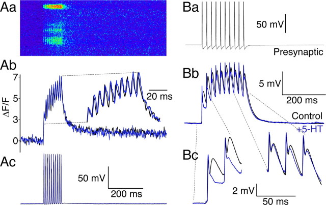
Presynaptic residual Ca2+ at presynaptic terminals coincides with a train-dependent loss of 5-HT-mediated presynaptic inhibition. A, Presynaptic Ca2+ transients were recorded by confocal line-scanning along the axonal plasma membrane at 500 Hz. Aa, Line-scan recording of Ca2+ dye fluorescence in axon (3 active zones). Axon labeled with low-affinity Ca2+ dye Fluo 5F from a recording microelectrode. Ten action potentials, 50 Hz, evoked summating Ca2+ transients. Ab, Ca2+ transients displayed as ΔF/F from uppermost active zone (control, black; 5-HT, 1 μm, blue). The time base of the traces is expanded in the inset to clearly demonstrate the absence of any effect of 5-HT. Ac, Presynaptic action potential train that caused the Ca2+ transients (control, black; 5-HT, 1 μm, blue) evoked by stimulation through the recording electrode. Aa–Ac are aligned and shown with the same time base. B, Current-clamp recording of train stimulation. Ba, Ten presynaptic action potentials, 50 Hz evoked through the presynaptic recording electrode. Bb, Average of 10 sequential postsynaptic traces in control (black) and in 5-HT (0.6 μm; gray). Note the effect of 5-HT is limited to the start of the train. Bc, Expanded recording of the first two and last three EPSPs in the train to emphasize the loss of inhibitory effect of 5-HT at the end of the train. The initial absolute voltage of the last three traces was adjusted to that of control to emphasize the later responses in the train were not significantly inhibited by 5-HT.

