Figure 6.
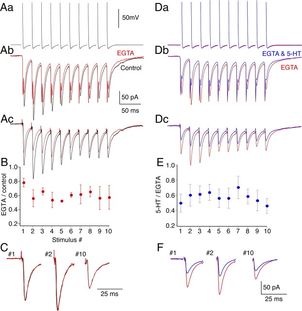
Train-dependent loss of 5-HT-mediated inhibition is prevented by presynaptic injection of EGTA. A, Paired recording between presynaptic reticulospinal axon and postsynaptic neuron in which EGTA (100 mm) was included in the presynaptic electrode. Aa, Presynaptic action potentials before (black) and after (red) pressure injection of EGTA into the axon. EGTA left action potentials unaffected throughout the train. Ab, Postsynaptic EPSCs comprising electrical and chemical components evoked by presynaptic action potentials. Traces are averages of 10 responses before and after injection of EGTA into the presynaptic axon. Ac, Chemical EPSCs after subtraction of the electrical components that were obtained after blocking AMPA-mediated responses with CNQX. B, Effect of EGTA injection as proportional inhibition for each of the 10 EPSCs in the train. Data are from four paired recordings and normalized to control amplitudes of each EPSC. The first response was inhibited to a lesser extent than later responses in the train. C, As representative examples, the first, second, and last EPSCs in the train were compared before and after EGTA injection. The responses after EGTA were scaled to the peak amplitudes of the control responses. The kinetics of the responses (both rise and decay) were unaltered. D, Paired recordings from cell in A; red traces are the same as those in A after presynaptic EGTA injection. Da, Presynaptic action potentials. Db, Evoked EPSCs in EGTA and 5-HT (600 nm; blue). Dc, Traces after subtraction of electrical components. E, Ratio of EPSC amplitudes in 5-HT to control (n = 5 preparations and pairs). 5-HT inhibited EPSCs throughout the train after presynaptic EGTA injection. Error bars indicate SEM. F, The first, second, and last traces are expanded to emphasize that inhibition is sustained throughout the train.

