
Figure 5.
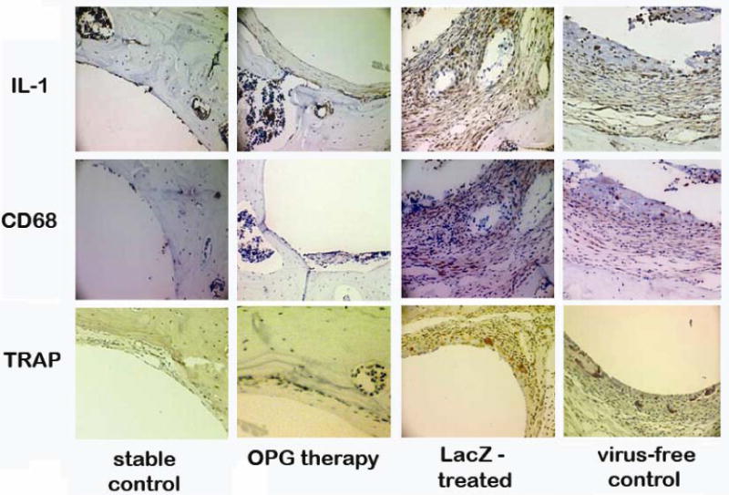
Typical IHC stained periprosthetic tissue sections against mouse IL-1 (upper panels), and mouse CD68 (middle panels) to reveal IL-1 expressing and CD68+ cells. TRAP staining to identify mature osteoclast at the bone-implant interface (lower panels). Four experimental groups are illustrated in columns.