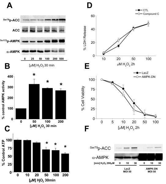
Figure 1. Peroxide induces AMPK activation in endothelium.
PAECs in 6-well plates were exposed to H2O2 as indicated, lysed, and the lysates probed for (A) phosphorylation of AMPK and ACC, (B) AMPK activity, and (C) ATP content as described in “Methods.” PAECs were then treated with H2O2 as indicated after treatment with either the AMPK inhibitor, compound C (25 uM; D) or dominant-negative AMPK adenovirus (E) and cell death or viability determined by LDH release and MTS assay, respectively as described in “Methods.” (F) PAECs were treated with dominant-negative AMPK adenovirus and AMPK activation assessed after H2O2 exposure by phosphorylation of the AMPK target, acetyl-CoA carboxylase (ACC).

