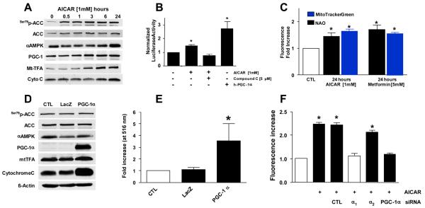
Figure 4. AMPK-activation in endothelial cells induces PGC-1α-dependent mitochondrial biogenesis.
(A) PAECs were exposed to AICAR as indicated, lysed, and subjected to immunoblotting for assessment of AMPK activation (p-ACC) and the levels of PGC-1α and mitochondrial transcription factor A (Mt-TFA). (A) BAECs were transfected with the Mt-TFA-promoter linked to a luciferase reporter31 prior to incubation with AICAR ± compound C followed by assessment of luciferase activity. Adenoviral transfection of human PGC-1α served as positive control. (C) PAECs were incubated with AICAR or metformin as indicated and mitochondrial mass determined fluorometrically with Mitotracker Green or nonyl-acridine orange (NAO) as indicated (*p<0.05 vs. CTL by two-way ANOVA and Dunnet's test) HUVECs were transfected with adenoviral vectors expressing either β-galactosidase (LacZ) or human PGC-1α. Cells were then lysed and assessed for the indicated proteins (D) or mitochondrial mass using Mitotracker (E; *p<0.05 vs. CTL by one-way ANOVA and a Dunnett's test). (F) HUVECs were treated with the indicated siRNA or buffer control for 72hr before a 24 hour incubation with AICAR. Mitochondrial mass was then determined using Mitotracker Green, *p<0.05 vs. no AICAR exposure. All experiments are N=5 – 7.

