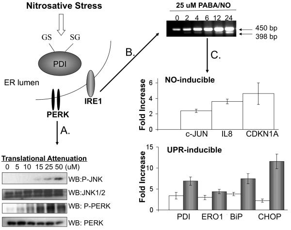
Figure 5. The mechanism of PABA/NO-induced antitumor activity is attributed to activation of the UPR.
The mechanistic studies were extended to HL60 cells in which S-glutathionylation of PDI was originally identified following PABA/NO treatment. HL60 cells were treated with 0-50 μM PABA/NO for 1h. Translational attenuation through activation of PERK was evaluated in HL60 cells following drug treatment (A). 50μg protein lysate was separated by SDS-PAGE and analyzed by immunoblot for levels of JNK and PERK. The membranes were stripped and probed for total levels of phosphorylated proteins. Activation of XBP-1 was detected by PCR (B). RNA was extracted at various time points and analyzed with primers specific to XPB-1. A 398 bp fragment corresponds to the cleaved and active form of XBP-1. Transcriptional activation of NO- and UPR-inducible proteins was evaluated with SYBR-green RT-PCR methods (C). HL60 cells treated with 25 μM PABA/NO for 4-6h (no shading) or 24h (gray shading). The relative quantity was measured as the ratio of the mRNA expression of the target gene to that of GAPDH. Data are the mean for 3 independent experiments +/− S.D.

