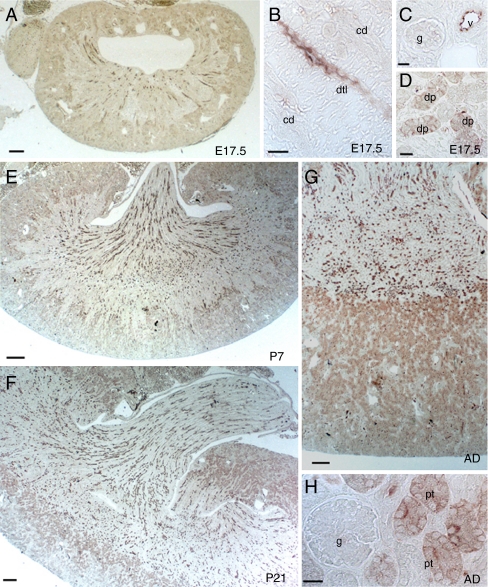
Fig. 6.
Segmental distribution of AQP1 during CD-1 mouse nephrogenesis. a–d Immunostaining for AQP1 (1:200) at E17.5. AQP1 immunoreactivity is detected in some medullary descending thin limb (dtl) and developing proximal tubules (dp) at E17.5. Glomeruli (g) and collecting duct (cd) are unstained. Luminal surface of blood vessels (v) are strongly stained. e–h. Postnatal increase in the density of AQP1-positive tubule profiles in the cortex and medulla at P7 (e), P21 (f), and in adult (g–h). Scale bars 200 µm

