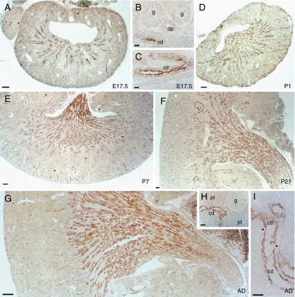
Fig. 7.
Segmental distribution of AQP2 during CD-1 mouse nephrogenesis. a–c Immunostaining for AQP2 (1:200) at E17.5. AQP2 immunoreactivity is detected in collecting ducts (cd). Glomeruli (g) and developing proximal tubules (dp) are negative. d–f Postnatal increase in AQP2 staining in the medulla, located in collecting ducts profiles at P1 (d), P7 (e), and P21 (f). g–i Adult kidneys with AQP2 staining in the collecting ducts (cd) (g, h) with negative proximal tubules (pt). Note the negative intercalated cells (asterisk) in the collecting ducts of mature kidney (i). Scale bars 200 µm

