
Figure 7.
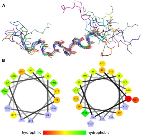
Structural model of warnericin RK in a membrane-like environment. (A) Superposition of the 10 lowest-energy structures calculated from the NMR data acquired in the presence of 8% TFE. (B) Helix wheel representation shows the amphiphilic character of warnericin RK residues 4–16 (left) and δ-lysin residues 2–21 (right). Hydrophobicity according to the color bar (charged residues are colored blue). Symbols: circles, hydrophilic; diamonds, hydrophobic; triangles, negatively charged; pentagons, positively charged.