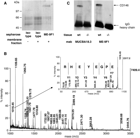
Fig. 1.
Preparation and identification of the ME-9F1 antigen. a SDS-PAGE analysis after immunoadsorption of solubilized membrane fractions of murine endothelioma cells with ME-9F1-Sepharose. Eluates of control-Sepharose and of control-Sepharose after incubation with membrane fraction (pre-adsorption) served as controls. b Peptide mass fingerprint of the SDS-PAGE separated ME-9F1 antigen by MALDI-MS. Inset: MS/MS spectrum of the peak with the mass of 1016.45. c Western Blot analysis of lung lysate from wild type or CD146−/− mice after staining with MUCBA18.3 or ME-9F1 and peroxidase anti-mouse IgG. The mouse IgG heavy chain served as equal loading control

