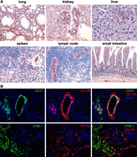
Fig. 2.
Tissue distribution of CD146 in murine organs. a Paraffin tissue sections (4 μm) were subsequently stained with the mab ME-9F1, biotinylated rabbit anti-rat antibody and streptavidine-alkaline phosphatase and developed with Fast Red, resulting in a red labeling of the endothelial cells of the blood vessels. Original magnification ×200. b Snap-frozen kidney tissue was stained for CD31 (green) and CD146 (red) (upper panel). Original magnification ×200. Paraffin tissue sections of lymph nodes were stained using a polyclonal antibody against LYVE-1 (green) and ME-9F1 against CD146 (red) (lower panel). Original magnification ×100. Nuclei were counterstained with DAPI (blue). Pictures representative for five mice studied independently

