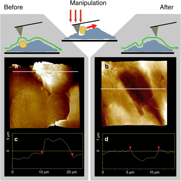
Fig. 3.

Nano-surgery at the docking site. Samples were prepared as in Fig. 2. a AFM contact image of a firmly attached leukocyte exhibiting a protrusion invading the endothelium. In a next run, increased force and high lateral speed were applied to tear out the leukocyte. b Repeated contact imaging of the identical area reveals a CLDS underneath. Here, it is located across a cell-junctional ridge. c, d The height profiles along the white lines in the images demonstrate how an 8-μm protrusion turns into an 1-μm invagination through distraction of the immune cell
