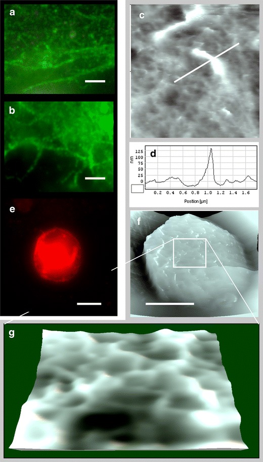
Fig. 5.

Human cell system. Freshly isolated blood neutrophils were added to primary human endothelial cells (HUVEC) grown on glass cover slips stimulated with TNFα. a, b Fluorescence micrographs of endothelial cells marked with antibodies against ICAM-1. Scale bar is 5 μm. Finger-like structures can be recognized that do not necessarily coincide with endothelial junctions. c AFM contact image of finger-like protrusions on an endothelial cell and d corresponding height profile. e Fluorescence micrograph of a leukocyte marked with antibodies against CD66. f, g AFM contact image of a neutrophil sitting on endothelium; g zoom into the top region reveals a sponge-like surface
