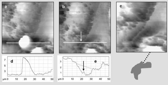
Fig. 6.

Segmented docking structure in human cells. A HUVEC/blood neutrophil cell sample underwent the nanomanipulation procedure as depicted in Fig. 3. a AFM image before and b after the removal of the leukocyte; c zoom into the docking region is illustrated by the sketch thereunder: the interface region is shaded brightly, the cup region in dark and the cell junction is given as a dotted line. In d and e, the corresponding height profiles are given with arrows denoting the ridge at endothelial cell junction
