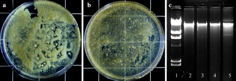
Fig. 3.
Growth of HP cells on solid MS medium established from control (a) and from cells co-cultivated for 5 days with A. tumefaciens (b) (pictures taken after 10 days of culture). c Agarose gel electrophorogram of HP genomic DNA isolated from control and cultures inoculated with bacteria after 3 days, lane 1HindIII digest of λ phage DNA, lane 2 control cells, lane 3 culture inoculated with A. tumefaciens, lane 4 culture inoculated with A. rhizogenes, lane 5 culture inoculated with E. coli

