
Figure 3.
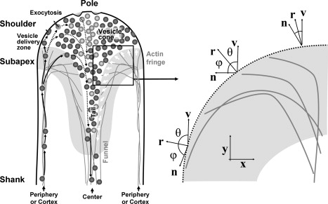
(Left) Schematic drawing illustrating the principal directions of vesicle motion (left half of the tube) and orientation of the actin filaments bordering the vesicle cone (open arrows in right half) in the apical region of a pollen tube. After delivery into the apical region on the actin filaments forming the fringe, vesicles are released into the apical cytoplasm in an annulus-shaped zone (vesicle delivery zone). Vesicles that succeed in contacting the plasma membrane undergo exocytosis. Vesicles that do not succeed in contacting the plasma membrane stream rearwards within the cone-shaped vesicle pool. Many of these vesicles are recirculated back into the forward stream immediately in the subapical region (not shown). Solid arrows indicate actin-myosin-guided vesicle movement, dashed arrows indicate movements that are presumably governed by diffusion. Objects are not drawn to scale. For clarity, except for vesicles, no other organelle or the cell wall is drawn. This figure is based on results by Bove et al. (1) and Zonia and Munnik (54). (Right) Orientation of the vectors along the actin fringe profile. The values r, n, and v are the vector normal to the profile, the microfilament orientation vector, and the growth vector of the cytoskeleton, respectively. The profile of the actin fringe is such that the angle ϕ between the normal vector and the actin microfilament orientation is equal to the angle θ between the normal vector and the growth direction, the y axis. Once the orientation vector n is fixed, the shape of the fringe profile can be determined.