
Figure 1.
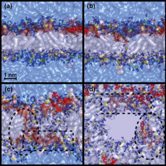
Four snapshots of an MD simulation. (a) Lateral view showing how the peptides are bound to the membrane before translocation. (b) Translocation of an Arg amino acid surrounded by water molecules that nucleates the formation of a water pore. (c) Lateral view of the pore, translocated peptide surrounded by a dotted square line, and a translocating peptide circled by an oval dotted line. (d) Top view of the pore. The phospholipid molecules are represented with transparent white surfaces, the phosphate atoms are in yellow spheres, the peptide molecules are in red, any water molecule at a distance of <0.35 nm from any phospholipids or amino acid atom is colored in solid blue, and the rest of the water molecules appear as a transparent blue surface.