
Figure 7.
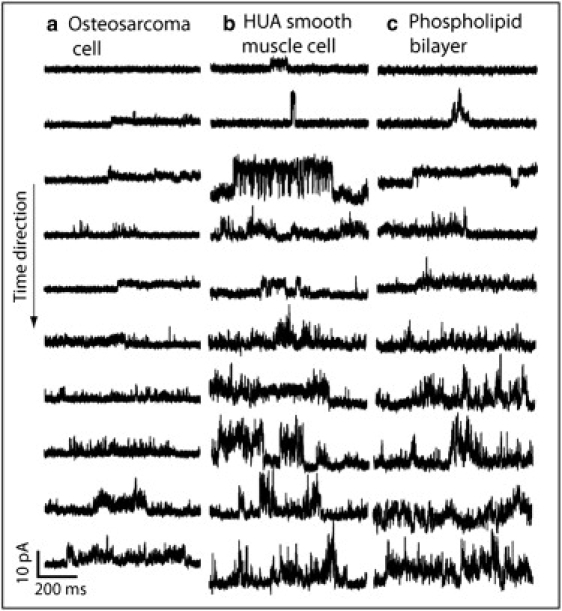
Snapshots of typical recordings of the current evoked by the peptide (placed inside the pipette) at different times after the seal was obtained in the cell-attached configuration in (a) an osteosarcoma cell, (b) an HUA smooth muscle cell, and (c) a phospholipid bilayer. It can be seen that as the time increases, the membrane is increasingly permeabilized, and the signal reflects a rapidly fluctuating current in every case.