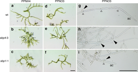
Fig. 2.
Microscopy of protoplast regeneration and side branch formation of ppsbp1 and ppsbp4 disruptants. Left and middle panels, regenerating protoplasts after 14 days on PPNH4 medium (a–c) and PPNO3 medium (d–f). Right panels, protonema grown on PPNO3 10 days after propagation (g–i). Regenerating protoplasts and protonema of wild type (wt, upper row) are shown for comparison. White triangles indicate anticlinal cell walls, black arrowheads side branches; ac apical cell. Bar represents 200 μm

