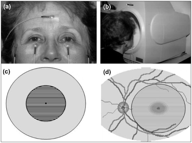
Figure 1. Setup for the non-invasive PERG.
(a) Standard skin electrodes are taped on the lower eyelids (active), temples (reference), and forehead (common ground). (b) The subject looks for 3 minutes at the center of the stimulus placed inside a ganzfeld bowl. (c) Pattern stimulus consisting of black and white stripes alternating 16.6 times per second. (d) The stimulus covers a circular retinal area with a 12.5 degree radius centered on the fovea.

