
Figure 3. In vivo GPR30 RNAi in the mediobasal hypothalamus.
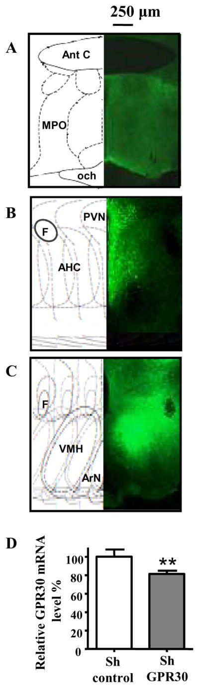
A to C: Typical spreading of virus expressing control or specific GPR30 shRNA, 3 to 6 weeks after bilateral injection of AAV1/2 into the mediobasal hypothalamus. Spreading of virus expressing eGFP was visible under the microscope by direct fluorescence visualisation. Fluorescence was seen at the level of the POA (A), paraventricular nucleus (B), and in the arcuate nucleus, median eminence and VMH (C).
D: Quantification of GPR30 in vivo knockdown. Animals were bilaterally injected with the control virus (shControl, n=12) or the virus expressing the specific shRNA for GPR30 (shGPR30, n=12) in the mediobasal hypothalamus. Five weeks later, GPR30 mRNA expression was measured by real time PCR using GAPDH as normalizer. The values in animals injected with the virus expressing shGPR30 are expressed relative to the values in animals receiving the control virus. **: P<0.005, T-test Ant C: Anterior commissure, MPO: medial preoptic area, och: optic chiasma, PVN:paraventricular nucleus, F: Fornix, AHC: Anterior Hyp. central area, VMH: Ventromedial hypothalamic nucleus, ArN: Arcuate nucleus.