
Figure 5.
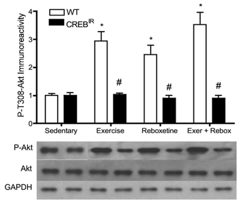
In WT mice, all interventions led to a significant activation of the signaling molecule, Akt, whereas no change from baseline was evident in CREBIR mice. *Among the WT mice, those who exercised (p < .0001, n = 27), received reboxetine (p = .027, n = 7), and received the combination of the two treatments (p < .0001, n = 7) expressed significantly more P-Akt immunoreactivity than those that were sedentary (p < .0001, n = 12). #WT mice also expressed significantly more P-Akt immunoreactivity than CREBIR mice for each intervention, except for sedentary mice, who expressed the same amount between genotypes. Among the CREBIR mice, there were no significant differences among treatments in P-Akt immunoreactivity. Because of the very small sample volumes, each mouse hippocampus was randomly assigned to a particular gel/Western film, thereby controlling for film-to-film variability. Therefore, the row of P-Akt bands and their respective Akt and GAPDH bands are a composite from several different Western blotting films. However, each P-Akt band and its respective Akt band and GAPDH band all represent the same mouse hippocampal sample (same gel lane).