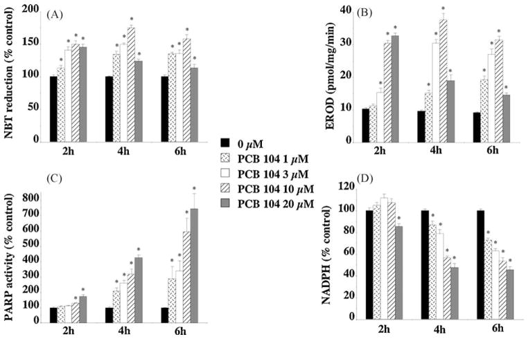
Fig. 3.
In vitro exposure of endothelial cells to PCB 104 dose- and time-dependently increased cellular levels of oxidative stress (A), CYP1A1 activity (B), and PARP activity (C) while depleting NADPH levels (D). Mouse endothelial cells were exposed to PCB 104 (1, 3, 10 or 20 μM) for 2, 4 or 6 h prior to the measurements being taken. Data is expressed as mean ± SEM from 4 separate experiments with 3–6 replicates per experiment; *p < 0.01 vs. untreated cells.

