
Figure 6. Neutralizing IL-6 reduces cardiac fibrosis.
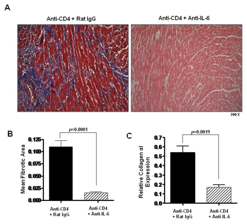
(A) Representative views of Masson’s trichrome staining of day 30 post transplant cardiac allografts taken from recipients transiently depleted of CD4+ cells which also received either control rat IgG antibodies (Anti-CD4 + Rat IgG) or neutralizing IL-6 mAb (Anti-CD4 + Anti-IL-6). (B) Morphometric analysis of graft fibrosis in groups described in (A). Bars represent the combined mean + S.E.M. of fibrotic (blue) area from at least 10 different frames of view from each of at least 6 different cardiac grafts. (C) Intragraft collagen αI message levels in cardiac allografts taken from recipients described in (B) were determined using quantitative real time PCR. Bars represent mean + S.E.M. of tissue harvested from at least 6 different transplants per group.