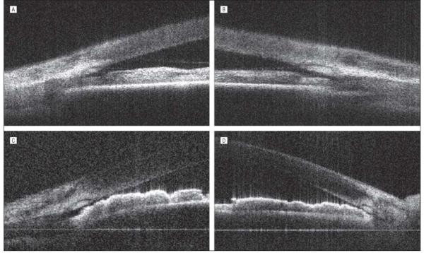
Figure 5.

High-resolution Fourier-domain optical coherence tomographic scans of narrow angles with gonioscopic grade I and grade 0. A, A very narrow space is visible between the root of the iris and the trabecular meshwork in the right eye of a patient with narrow-angle glaucoma (gonioscopic grade I). B, The left eye of the same patient (A) shows similar findings. C, Near touch between the trabecular meshwork and the root of the iris is visible in the right eye of another patient with narrow-angle glaucoma (gonioscopic grade 0). D, The left eye of the same patient (C) shows a similar angle configuration.
