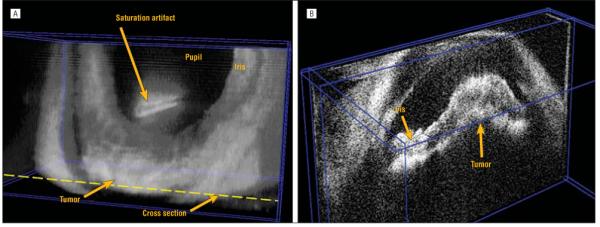
Figure 9.

An eye with a ciliary body tumor. A, A volumetric rendering showing irregularity of the anterior chamber depth due to elevation of the peripheral iris in the inferior region of the eye. B, A cross-sectional plane through the tumor location. Note that the actual tumor of the ciliary body is not visible.
