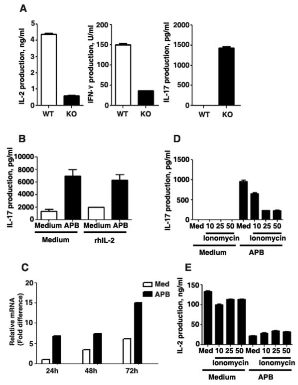
Fig. 5. IP3R-mediated Ca++ release suppresses IL-17 expression.
(A) CD4 T cells from wildtype (WT) or Ets-1 deficient (KO) mice were activated with anti-CD3/anti-CD28 mAbs and supernatant was analyzed for IL-2, IFNγ and IL-17 production by ELISA. (B) CD4 T cells were activated with anti-CD3/anti-CD28 mAbs for 4 days in the presence or absence of 2-APB (15 μM). Medium or rhIL-2 (100 ng/ml) was added to culture on day 0 of activation. On day 4, supernatant was analyzed for IL-17 production. (C) CD4 T cells were activated with anti-CD3/anti-CD28 mAbs for 1, 2 or 3 days in the presence or absence of 2-APB (15 μM). Relative mRNA levels of IL-17 were examined by real-time RT-PCR. (D & E) CD4 T cells were activated with anti-CD3/anti-CD28 mAbs in the presence or absence of 2-APB for 2 or 4 days. Ionomycin was added at 10, 25 or 50 ng/ml concentrations to control or 2-APB treated cells at the time of activation. Supernatants were analyzed for IL-17 production (D) on day 4 or IL-2 production (E) on day 2.

