
Figure 3.
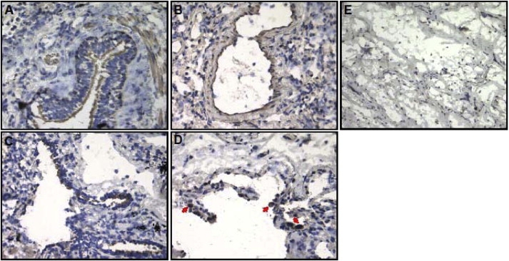
IREB2 Localization in Normal and Emphysematous Human Lung Tissue
(A–D) IREB2 protein expression by DBA staining (brown) and counterstaining by nuclei staining (blue). IREB2 expression in control tissue localized to (A) bronchial epithelial cells and (B) vascular endothelial cells; IREB2 expression in emphysematous tissue localized to (C) bronchial epithelial cells and (D) macrophages (red arrows).
(E) Negative control without primary antibody in control tissue. Magnification is 200×.