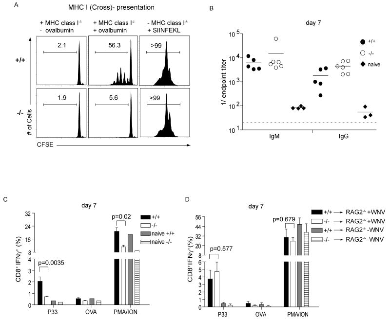
Fig. 3. Lack of cross-presentation and antiviral CTL responses in Batf3-/- mice.
(A) Batf3+/+ (+/+) or Batf3-/- (-/-) splenocytes were depleted of B220+ B cells and Thy1.2+ T cells and enriched for CD11c by MACS and cultured with irradiated MHC-class I-/- splenocytes as indicated that were either untreated (-ovalbumin), pulsed with 10 mg/ml soluble ovalbumin (+ovalbumin), or cultured with 1 μM SIINFEKL peptide. CFSE-labeled CD45.1+ OT-I T cells were cultured with these cells and proliferation determined by FACS after 60 hours. Single-color histograms of CD8+CD45.1+ OT-I T cells show the percentage of cells in the indicated gates. (B) Batf3+/+ (+/+) or Batf3-/- (-/-) mice were infected with 100 PFU of WNV. On day 7, isotype-specific anti-WNV E protein titers were measured. (C) Batf3+/+ (+/+) or Batf3-/- (-/-) mice were infected with 100 PFU of WNV, or left uninfected. After 7 days, splenocytes were stimulated in vitro with the WNV-specific NS4B peptide (P33), OVA peptide, or PMA/ionomycin as described. CD8+ T cells were analyzed for expression of intracellular IFN-γ. Data shown are the mean ± SEM (n=9-10). (D) Batf3+/+ (+/+) or Batf3-/- (-/-) CD8+ T cells were transferred i.v. into Rag2-/- recipients. After 24h, mice were infected with 100 PFU of WNV (+ WNV) or left uninfected (-WNV). After 7 days, splenocytes were harvested and analyzed as described in (C). Data shown are the mean ± SEM (n=6). Three independently performed experiments yielded similar results.

