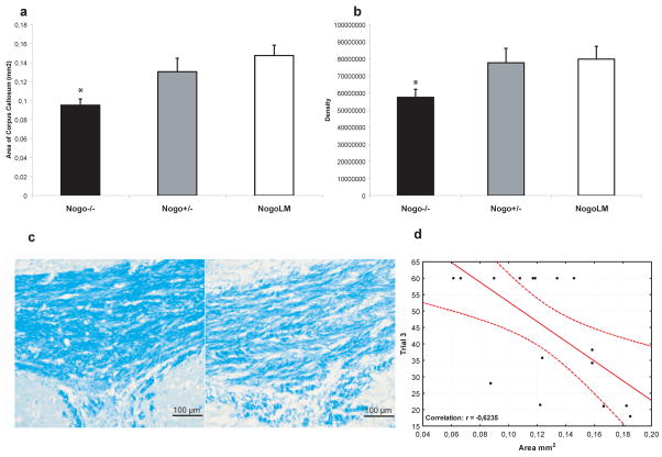
Fig. 5.
We evaluated myelin staining in the corpus callosum (CC) of brain-injured animals. We observed that brain-injured Nogo-A−/− mice had significantly (*p<0.05) reduced area of the CC (a) and in the CC, reduced myelin density (b) measured in arbitrary units (pixels/area unit) compared to their brain-injured, wild-type littermates. In (c), representative Images of the CC of a littermate, brain-injured control (left) and a NogoA/B−/−, brain-injured animal (right) is shown. Scale bar =100 μm. MWM performance (shown for trial block 3) was negatively correlated with CC area (d).

