
Figure 7.
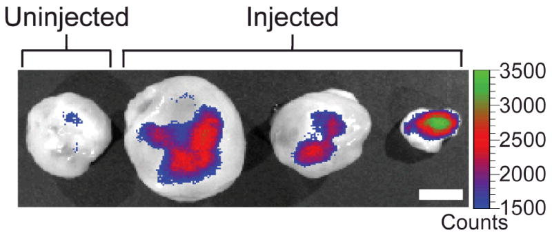
Xenogen IVIS fluorescence images of flank xenograft C6 tumors of different sizes excised from three mice injected with NP-CP-PEI:DNA and a mouse receiving no injection. The scale bar corresponds to 5 mm.
Official websites use .gov
A
.gov website belongs to an official
government organization in the United States.
Secure .gov websites use HTTPS
A lock (
) or https:// means you've safely
connected to the .gov website. Share sensitive
information only on official, secure websites.
Xenogen IVIS fluorescence images of flank xenograft C6 tumors of different sizes excised from three mice injected with NP-CP-PEI:DNA and a mouse receiving no injection. The scale bar corresponds to 5 mm.