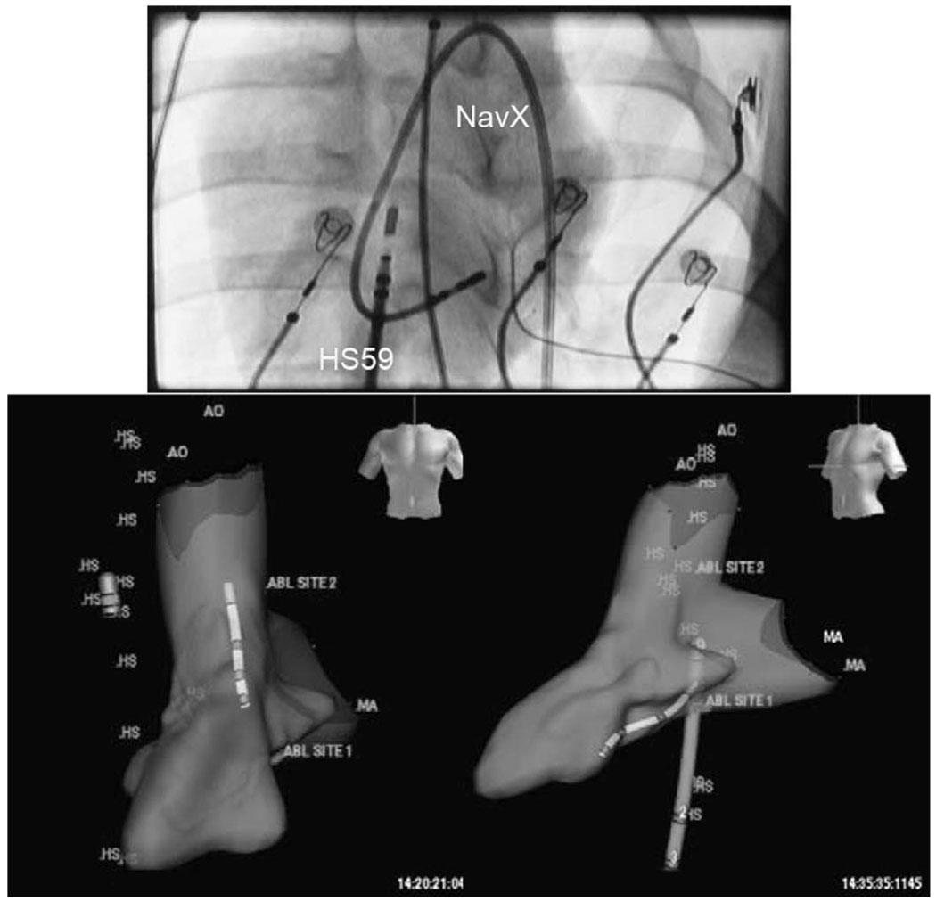
Fig. 13.
The anterior view fluoroscopic image at the top shows the NavX mapping catheter looped on itself in the left ventricle (LV) of a pig, while the HockeyStick is shown in the RA. The two views at the bottom show the 3-D volume of a pig LV mapped by a NavX electroanatomical mapping catheter; the HockeyStick catheter (labeled “HS”) is in the RA while the slightly smaller NavX catheter is seen in the left ventricle. Both catheters were independently steered and could be tracked in a continuous 3-D mode very easily.

