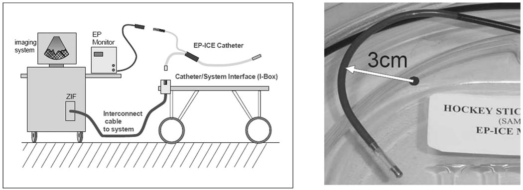
Fig. 2.
The general cable connection scheme for the combination catheter. The left panel shows the nondisposable trunk cable between the imaging system and the patient table, and the two separate connection paths for EP mapping and for imaging. The right panel shows the distal end of an early mechanical prototype steered in its minimum bend radius position.

