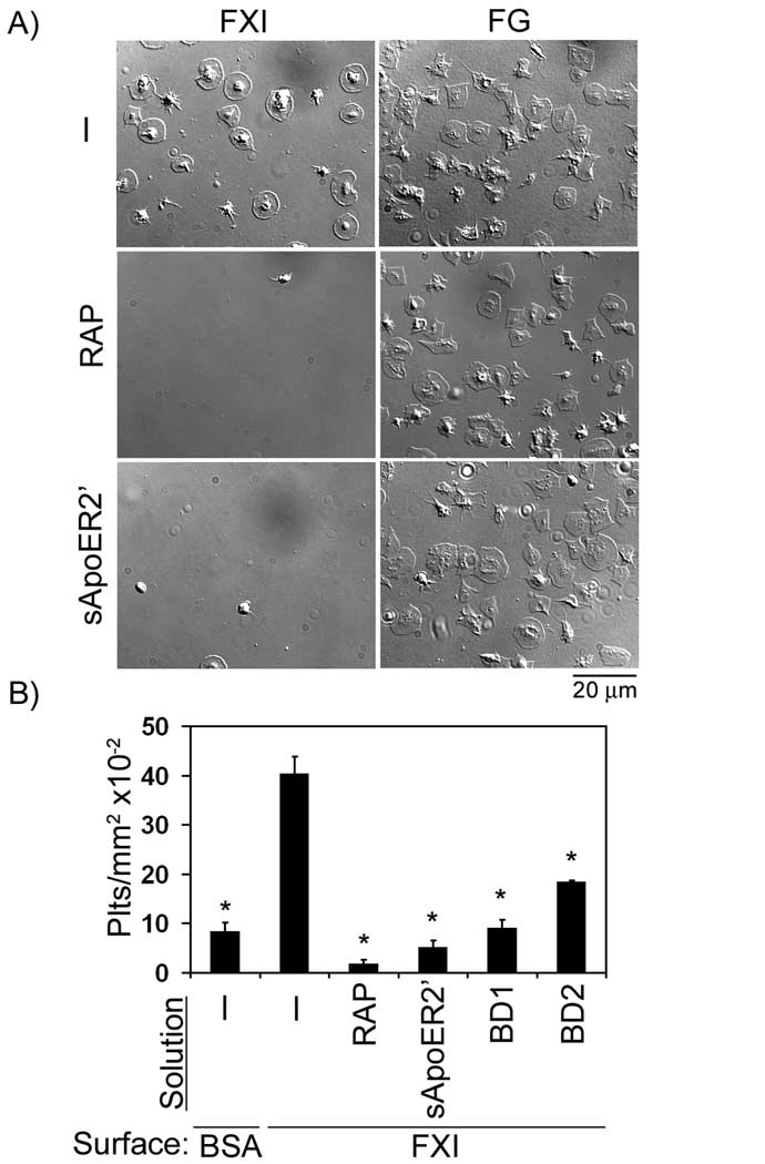
Figure 2. The role of ApoER2 in platelet adhesion to FXI.
(A) Purified human platelets (2 × 10⁷/ml) were pipetted onto surfaces coated with FXI or fibrinogen (FG) in the absence or presence of receptor-associated protein (RAP, 40 µg/ml) or soluble ApoER2’ (sApoER2’, 40 µg/ml), followed by incubation at 37°C for 45 min. (B) Additional adhesion experiments were performed on immobilized FXI in the presence of the LDL binding domains 1 or 2 of ApoER2 (BD1 or BD2, respectively, 40 µg/ml). Adherent platelets were analyzed for each suspension treatment and reported as adherent cells/mm2 × 10−2. Values are reported as mean ± SEM of three experiments. *P < 0.05 compared to adhesion on FXI in the presence of vehicle.

