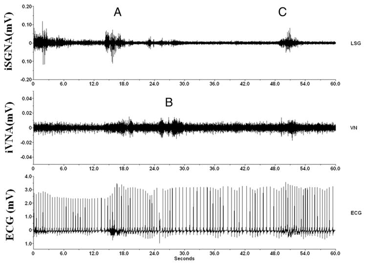
Figure 1. Nerve and ECG Recordings.
Example of simultaneous integrated stellate ganglion nerve activity (iSGNA) and integrated vagal nerve activity (iVNA) at baseline (before pacing-induced congestive heart failure) in an ambulatory dog. (Top) iSGNA; (middle) iVNA; (bottom) electrocardiographic (ECG) trace. In the upper trace, A indicates between the 12th and 16th s, an epoch of high sympathetic activity followed 3 s later by an epoch of fast heart rate (about 120 beats/min, between the 18th and 24th s). This sympathetic activity is followed almost immediately, between the 24th and 30th s, by a vagal increase with lower heart rate (B) (from 120 to 90 beats/min, between the 30th and 36th s), and last (C), a new increase in sympathetic activity after the 48th s, and a heart rate increase (from 90 to 130 beats/min, around the 54th s). LSG = left stellate ganglion nerve; VN = vagal nerve.

