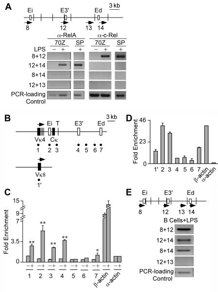
FIGURE 6.
Potential RelA, c-Rel and RNA polymerase II interactions bridging the Igκ gene enhancers. A, ChIP-3C assays of RelA’s and c-Rel’s presence in Ei-E3′, E3′-Ed, and Ei-Ed complexes in 20 h LPS treated 70Z/3 cells and 3 day LPS stimulated splenic B cells (SP). Pre-immune IgG immunoprecipitation controls reveal that no amplification products are detected. B, Map of the Igκ locus with the sites assayed for by PCR amplification in ChIP samples indicated by the underlying numbered solid circles. C, Real time PCR ChIP assays of Pol II occupancy at the Igκ locus in 70Z/3 cells before (-) and after 20 h of LPS treatment (+). Fold enrichment is defined in Figure 5G. Standard deviations of three independent chromatin preparations are indicated. D Real time PCR ChIP assays of Pol II occupancy at the Igκ locus in splenic B cells after 3 days of LPS treatment. Fold enrichment is defined in Figure 5G. Standard deviations of three independent chromatin preparations are indicated. E, ChIP-3C assay for presence of Pol II in the Ei-E3′, E3′-Ed, Ei-Ed complexes in splenic B cells stimulated with LPS for 3 days. Pre-immune IgG immunoprecipitation controls reveal that no amplification products are detected.

