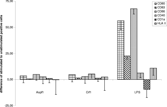
FIG. 4.
Expression of selected DC markers after stimulation with Aspf1, Crf1, and LPS by flow cytometric analysis of selected DC surface markers (CD1a, CD40, CD80, CD83, CD86, HLA class II). DCs were stimulated for 48 h with Aspf1, Crf1, and LPS, and their expression (in percent) was compared to that of unstimulated DCs.

