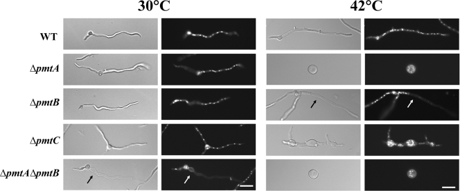
FIG. 4.
Phenotypes of Δpmt mutants. Conidia of Δpmt mutants were inoculated onto CM, incubated for 12 h at 30°C or 42°C, fixed, and stained with Hoechst and Calcofluor white to label the nuclei and cell walls, respectively. (Left) Differential interference contrast images. (Right) Fluorescence images. The arrow indicates an empty apical compartment. Bar, 10 μm. WT, wild type.

